Not all of us are medical gurus. Most who understand all the fancy lingo that goes into explaining what certain procedures and practices entail are either doctors, or someone who’s been diagnosed with a certain disease. The latter will have spent hours scouring the Internet for answers to what ails them, on sites like: WebMD, Mayo Clinic, the CDC and Drugs.com. Once you’ve been diagnosed — self or real — you’ll need to figure out your course of treatment, which may include X-rays, MRIs, CT scans or a combination of these digital imaging practices.
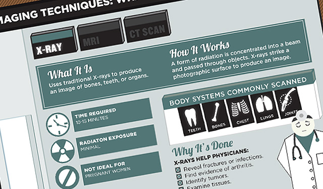
The problem with being a member of the layperson masses, is that once you figure out what ails you, what are the procedures to getting healthy again? Will you need digital imaging to find out what’s going wrong with your insides, and if you do, which technique is the one for you? This infographic created by Concordia University St Paul will help you figure out all your questions about digital imaging.
X-Rays
Traditional X-rays are used to produce an image of the bones, tech, or organs. They work by forming using a form of radiation that is concentrated into a beam and passed through objects. These X-rays strike a photographic surface to produce an image.
MRIs
An MRI machine uses a strong magnet to produce an image of organs, tissue, and skeletal systems. It’s used mainly to identify injury, bleeding, tumors, infection, or injury. It works by placing the patient inside a specialized machine that uses a magnetic field and radio wave energy to produce images of organs and internal structures.
CT Scans
CT Scans use X-rays to create a digital 3D or 4D image of organs, tissue, and other body structures. It works by sending an X-ray pulses through the body to create images of thin slices of the area. The images are then used in a series to create a more detailed image.
Check out the graphic below to see which body system each different form of digital imaging is most commonly used on.

